
Fig. 3
- ID
- ZDB-FIG-150511-10
- Publication
- Otis et al., 2015 - Zebrafish as a model for apolipoprotein biology: Comprehensive expression analysis and a role for ApoA-IV in regulating food intake
- Other Figures
- All Figure Page
- Back to All Figure Page
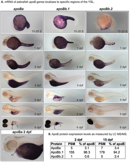
Zebrafish apoB mRNA and protein expression. (A) Localization of apoBa, apoBb.1 and apoBb.2 mRNA by ISH from somitogenesis [15-20 somite (S)] to 6 dpf. All apoB genes are expressed in the yolk syncytial layer (YSL), but mRNAs localize to distinct subregions of the YSL at various developmental stages. apoBa mRNA is present in the heart (H) at 2 dpf. At 6 dpf apoBa mRNA is expressed strongly in the liver (L) and weakly in the intestine (I), apoBb.1 is strongly expressed in the intestine and weakly in the liver, and apoBb.2 is not detectable by ISH. All zebrafish are wild type except 6-dpf larvae which are nacre-/-. Larvae collected at 2-5 dpf were treated with PTU to prevent pigment formation. No signal was observed at the eight-cell stage or at 30% or 80-100% epiboly (supplementary material Fig. S2). Experiments were performed three times for each gene at each stage with ne5 embryos or larvae per probe per experiment. (B) Relative abundance of zebrafish ApoB paralogs as identified by peptide spectrum matches (PSM) in 2- and 15-dpf larvae (20-30 pooled larvae per experiment) as determined by LC-MS/MS of total larval proteins ≥250 kDa. Two experiments were performed for each developmental stage and both experiments yielded similar results. Data presented are from one experiment. |
| Genes: | |
|---|---|
| Fish: | |
| Anatomical Terms: | |
| Stage Range: | 14-19 somites to Day 6 |