
Fig. 1
- ID
- ZDB-FIG-150430-27
- Publication
- Akiyama et al., 2014 - An anterior limit of FGF/Erk signal activity marks the earliest future somite boundary in zebrafish
- Other Figures
- All Figure Page
- Back to All Figure Page
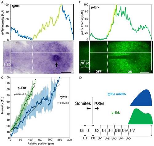
fgf8a mRNA shows graded distribution in the PSM, whereas FGF downstream signal activity, represented by p-Erk levels, does not. (A,B) Representative images of fgf8a expression (A) and p-Erk distribution (B) showing a gentle slope and a steep gradient, respectively. Signal intensities (graphs; AU, arbitrary unit) were measured by ImageJ software from the original images. Anterior and posterior ends of the PSM are marked by white lines. The position of the anterior extremity (dotted lines) was estimated by comparison of signal intensity (upper panel) and visual observation (lower panel). Dorsal view of tailbud regions in flat-mounted embryos, anterior to the left. The horizontal white lines in the lower images mark the paths along which the signal intensities shown in the upper panel were recorded. Arrow indicates Kupffer’s vesicle. Scale bars: 100 µm. (C) Comparison of slopes between fgf8a expression and p-Erk distribution. Intensity plots of the upward slope regions of the gradients represented by the yellow line in the graphs in A and B. Approximation formulae of fgf8a (n=8) and p-Erk (n=7) were calculated from average values of signal intensities, respectively. Dots indicate averages and green/blue bars indicate s.d. (D) Schematic of the distribution of fgf8a mRNA (blue) and p-Erk (green). B, boundary; PSM, presomitic mesoderm; S, somite. Prospective somites and future somite boundaries in the PSM are numbered as described by Pourquié and Tam (Pourquié and Tam, 2001). |
| Gene: | |
|---|---|
| Antibody: | |
| Fish: | |
| Anatomical Terms: | |
| Stage: | 5-9 somites |