
Fig. 1
- ID
- ZDB-FIG-150428-8
- Publication
- Luz et al., 2013 - Fluorescently tagged Lin7c is a dynamic marker for polarity maturation in the zebrafish retinal epithelium
- Other Figures
- All Figure Page
- Back to All Figure Page
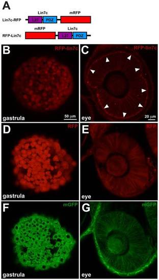
Lin7 sub-cellular localization in neuroepithelial cells is distinct from gastrula cells. Confocal live imaging of RFP-Lin7 sub-cellular localization in zebrafish embryos. (A) Schematic representation of Lin7 fusion proteins with monomeric RFP (mRFP). (B,D,F) In gastrula embryos (7 hpf, 70% epiboly stage) RFP-Lin7c, RFP and mGFP are distributed homogeneously in the cells. RFP-Lin7c and RFP are also detected in the nucleus. Scale bar: 50µm. (C,E,G) Retinal neuroepithelium at 28 hpf. RFP-Lin7c localizes to the cell membranes and is particularly enriched at the apical surface (arrowheads, C). RFP is distributed homogeneously in the cytosol (E) and mGFP localizes to the cell membranes, but is not enriched at the apical surface (G). Scale bar: 20 µm. |