
- Title
-
Fluorescently tagged Lin7c is a dynamic marker for polarity maturation in the zebrafish retinal epithelium
- Authors
- Luz, M., and Knust, E.
- Source
- Full text @ Biol. Open
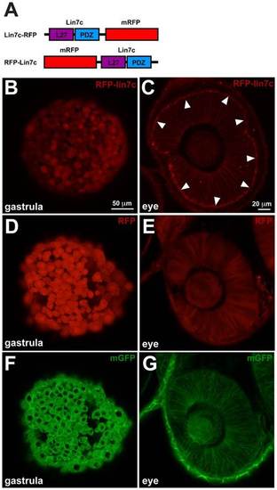
Lin7 sub-cellular localization in neuroepithelial cells is distinct from gastrula cells. Confocal live imaging of RFP-Lin7 sub-cellular localization in zebrafish embryos. (A) Schematic representation of Lin7 fusion proteins with monomeric RFP (mRFP). (B,D,F) In gastrula embryos (7 hpf, 70% epiboly stage) RFP-Lin7c, RFP and mGFP are distributed homogeneously in the cells. RFP-Lin7c and RFP are also detected in the nucleus. Scale bar: 50µm. (C,E,G) Retinal neuroepithelium at 28 hpf. RFP-Lin7c localizes to the cell membranes and is particularly enriched at the apical surface (arrowheads, C). RFP is distributed homogeneously in the cytosol (E) and mGFP localizes to the cell membranes, but is not enriched at the apical surface (G). Scale bar: 20 µm. |
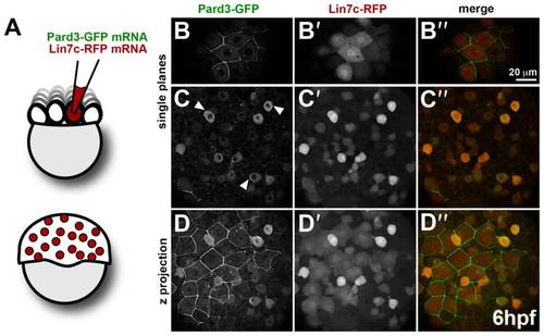
Comparison of Pard3-GFP and Lin7c-RFP subcellular localization reflects tissue-specific differences in epithelial polarity. (A) Schematic representation of mRNA injection in 1/16–32 blastomeres for mosaic expression. (B–D′′) Mosaic co-expression of Par3-GFP and Lin7c-RFP in the gastrula (6 hpf). Par3-GFP is restricted to the membrane of the enveloping layer (EVL) cells (B), while it is mainly cytosolic in the deep cell layer (DEL), with some local accumulations at the cortex (arrowheads) (C). Lin7c-RFP is cytosolic and nuclear both in the EVL (B′) and DEL (C′). (D–D′′) Maximum projection of the embryo showing Pard3-GFP (D), Lin7c-RFP (D′) and the merge of both (D′′) in the EVL and DEL. Scale bar: 20 µm. Single confocal planes are shown, except for D–D′′. |
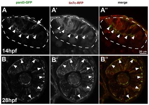
Lin7c-RFP subcellular localisation reflects temporal changes in retinal neuroepithelial polarity. (A–A′′) In the optic vesicle (14 hpf, 10ss), Pard3-GFP is observed predominantly at the apical surface (arrowheads) of cells, except for a few cells, which show a cytoplasmic distribution (arrow) (A). Lin7c-RFP is cytosolic and not observed apically (A′). (B–B′′) Cells of the retinal neuroepithelium (28 hpf) show apical localization of both Pard3-GFP (B) and Lin7c-RFP (B′). Scale bar: 20µm. |
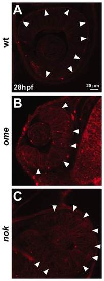
Nok and Ome are required for apical localisation of RFP-Lin7c. RFP-Lin7c sub-cellular localisation in the retinal neuroepithelium at 28 hpf. (A) In wild-type (wt) embryos, RFP-Lin7c is associated with the cell membranes, with a strong accumulation at the apical side (arrowheads). (B,C) RFP-Lin7c apical localisation is lost in ome and nok mutants (arrowheads). Note that in both mutants membrane association of RFP-Lin7c is maintained. Scale bar: 20 µm. |