
Fig. 3
- ID
- ZDB-FIG-150422-33
- Publication
- Stooke-Vaughan et al., 2015 - Otolith tethering in the zebrafish otic vesicle requires Otogelin and α-Tectorin
- Other Figures
- All Figure Page
- Back to All Figure Page
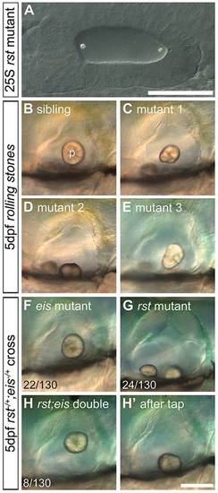
Development of otoliths in rst mutant and rst;eis double-mutant embryos. Live DIC micrographs of sibling and rst mutant OVs. The left OV is shown, with anterior to the left and dorsal to the top. (A) 25S embryo from a homozygous mutant rst cross. The otoliths seed normally at the anterior and posterior poles (compare with Fig. 1A). (B) 5dpf phenotypically wild-type sibling OV. p, posterior otolith. (C) 5dpf rst mutant OV. The posterior otolith is misshapen, detached and free to move. (D) OV of a different rst mutant, with a posterior otolith that has become detached and stuck on the ventral floor of the OV. (E) OV of an rst mutant with the most common phenotype of a misshapen posterior otolith. (F) 5dpf eis mutant with a single otolith tethered to the posterior macula. (G) 5dpf rst mutant with an untethered posterior otolith. (H) 5dpf rst;eis double mutant with a single untethered otolith. (H′) The same embryo as in H, after the slide was tapped to displace the untethered otolith. Numbers indicate embryos showing each phenotype (detached or misshapen posterior otolith) among total examined (130) from an rst;eis double-heterozygote cross; the remaining 76/130 embryos were phenotypically wild type (not shown). Scale bars: 50µm in A; 100µm in B-H′. |
| Fish: | |
|---|---|
| Observed In: | |
| Stage Range: | 20-25 somites to Day 5 |