
Fig. 5
- ID
- ZDB-FIG-150326-46
- Publication
- Skaggs et al., 2014 - Excitotoxic brain injury in adult zebrafish stimulates neurogenesis and long-distance neuronal integration
- Other Figures
- All Figure Page
- Back to All Figure Page
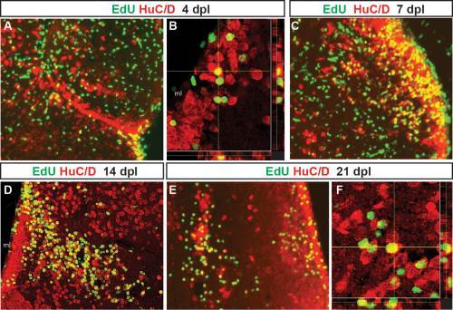
Adult-generated cells differentiate into neurons that progressively migrate to the site of injury. Proliferating cells that incorporated EdU at 2 days post-lesion were double labeled for EdU (green) and the neuronal marker HuC/D (red) after different chase periods. A yellow asterisk denotes the injury site in each panel. At 4 days after QA injection, EdU+ cells that express the neuronal marker HuC/D appeared to leave the VZ and extend toward the site of injury (A, orthogonal projection in B). By 7 dpl (C), EdU/HuC/D colabeled cells were found at a distance from the VZ. By 14 (D) and 21 dpl (E, orthogonal projection in F), two distinct populations of newborn neurons were present. Many double-labeled cells settled along the periventricular region where neuronal cell bodies are normally located. Other EdU/HuC/D double-labeled cells were located near the lesion site. Scale bars = 100 µm. |