|
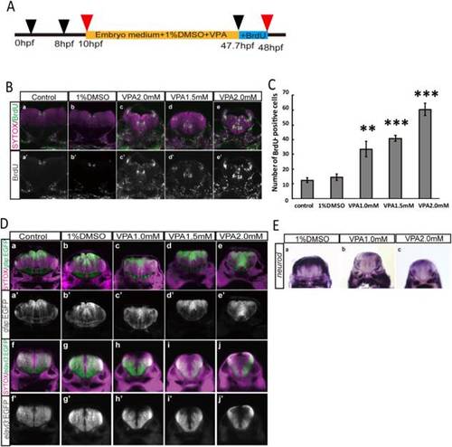
VPA increases proliferating cells in the hindbrain of embryonic zebrafish. A: Embryonic zebrafish were treated with 1.0, 1.5, 2.0 mM VPA from 10 to 48 hpf. In last 20 min, BrdU was added to label proliferating cells. B: Number of BrdU-positive cells increased in a dose-dependent manner, and the area of BrdU-positive cells expanded in the VPA-treated brain. C: Number of BrdU-positive cells per section. D: Tg(elavl3:GFP) and Tg(gfap:GFP) were treated with VPA at indicated concentrations, and the GFP-positive area was analyzed. Reduction of elavl3-GFP-positive area (a-e, a′-e′) and increase of gfap-GFP-positive area (f-j, f′-j′) were observed in VPA-treated zebrafish hindbrain. E: In situ hybridization of VPA-treated embryonic brain with neurod probes. Area of neurod-positive decreased in VPA-treated zebrafish hindbrain. After whole-mount in situ hybridization, coronal sections were made at rhombomere 5 or 6 of the hindbrain.
|