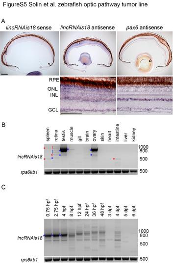
Examination of zebrafish lincRNAis18 expression in early development and adult tissues. (A) Nested RT-PCR showing expression of lincRNAis18 within the adult zebrafish retina. (B) In situ hybridization using non-radioactive DIG-labeled lincRNAis18 probes on adult zebrafish retina cryosections. lincRNAis18 expression is detected in the ganglion cell layer (GCL) and a subset of cells at the vitreal side of the inner nuclear layer (INL) (left, middle). Negative control, lincRNAis18 sense DIG-labeled probe (right). (C, D) RT-PCR showing relative levels of lincRNAis18 expression throughout development and in adult tissues. Control, expression of ribosomal protein S6 kinase b, polypeptide 1, rps6kb1. Blue bracket and asterisks indicate lincRNAis18 cloned and sequence confirmed products. Red bracket and asterisks indicate nonspecific products cloned and confirmed by sequencing. GCL, ganglion cell layer; INL, inner nuclear layer; ONL, outer nuclear layer; RPE, retinal pigmented epithelium. Scale bars 100 µm.
|
