|
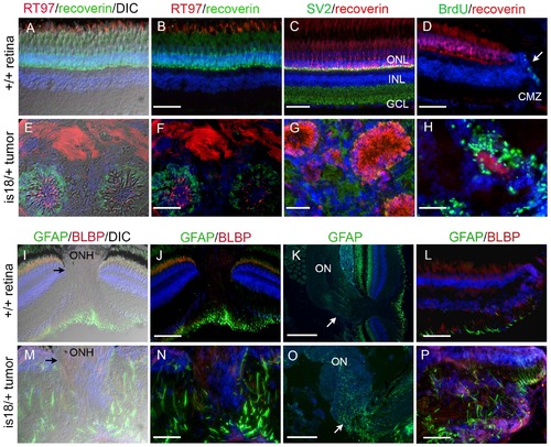
Characterization of lincRNAis18 tumors indicates a glial cell origin. Immunolabeling and in situ hybridization of cryosections from wild type retina (A–D, I, J, M–P) and advanced lincRNAis18 tumors (E–H, K, L, Q–T). Cells in rosettes in the tumors label with neurofilament marker RT-97 (red) and photoreceptor marker recoverin (green) (E, F). The synaptic vesicle marker SV2 (green), which is enriched in the retinal plexiform layers (C), was distributed throughout the fibrous tumor mass (G). (D) BrdU (green) incorporated into proliferating progenitor cells at the ciliary marginal zone of the wild type retina (arrow). (H) Intense labeling of BrdU incorporation was detected in cells forming rosettes and throughout the tumor mass. In wild type the glial marker GFAP is most evident in the Müller glia end feet that sit in the retinal ganglion cell layer (I–L). GFAP expression is absent in the oligodendrocytes and astrocytes of the optic nerve (I, K arrow). In contrast, GFAP was readily detected in long streaks throughout the tumor tissue (M, N, P), and was expressed by cells in the mutant optic nerve (O, arrow). BLBP is present at the ciliary marginal zone of wild type retina (L) and appeared present throughout the tumor mass (P). A, E, I, M, Differential interference contrast (DIC) overlay on immunofluorescence labeling images. CMZ, ciliary marginal zone; GCL, ganglion cell layer; INL, inner nuclear layer; ON, optic nerve; ONH, optic nerve head; ONL, outer nuclear layer. All scale bars represent 50 µm, except in panels K and O scale bars represent 100 µm.
|