
Fig. 1
- ID
- ZDB-FIG-150313-22
- Publication
- De Smet et al., 2014 - Fibroblast Growth Factor Signaling Affects Vascular Outgrowth and Is Required for the Maintenance of Blood Vessel Integrity
- Other Figures
- All Figure Page
- Back to All Figure Page
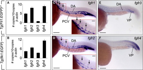
FGFR Expression in Zebrafish Embryos (A and B) Endothelial expression of FGFRs was determined by real-time RT-PCR analysis of GFP+ ECs isolated by flow cytometry from 24 hpf Tg(fli1:EGFP)y1 and Tg(flk1:EGFP) zebrafish embryos. Data are mean ± SEM. (C–F) Whole-mount in situ hybridization of 24 hpf embryos revealed that fgfr1 and fgfr2 were expressed in the DA, PCV, intersegmental vessels (ISVs; arrows) and vascular plexus (VP) in the ventrocaudal tail, while expression of fgfr3 in these vessels was less pronounced. The inset in C and D displays a higher magnification of the fgfr1- and fgfr2-positive ISVs (red boxed ares). Fgfr3 and fgfr4 are both present in the vascular plexus (VP), while fgfr3 is faintly expressed in the DA, and fgfr4 is expressed in the caudal somites. Fgfr4 was mainly present in the vacular plexus at 24 hpf. Scale bars denote 100 µm in (C)–(F). |
| Genes: | |
|---|---|
| Fish: | |
| Anatomical Terms: | |
| Stage: | Prim-5 |