
Fig. 3
- ID
- ZDB-FIG-150312-49
- Publication
- Preuss et al., 2014 - Classification of Object Size in Retinotectal Microcircuits
- Other Figures
- All Figure Page
- Back to All Figure Page
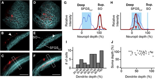
SINs Stratify in Different Sublayers of Retinorecipient Neuropil (A) Maximum-intensity projection showing a SIN filled with sulforhodamine (red) and retinal afferent fibers (cyan). A dorsal view is shown. (B) Transverse optical section through same cell as in (A). Dendrites of labeled SIN colocalize with the superficial SO band (arrowhead). (C) Maximum-intensity projection along same orientation as in (B). Dendrites remain in the SO layer. Note that the apparent broadening of the SIN dendrites (lower left) is caused by the tectal curvature. Compare also Movie S1. (D–F) Representation of a patched SIN in a different fish. The same orientation as in (A)–(C) is used. Note that dendritic tree stratifies in a narrow region of the deeper SFGSD,F. (G) Line profile analysis from boxed region in (C). The blue curve indicates GFP-expressing layers in SO and SFGSD,F. The red curve indicates the extent of the SIN dendritic tree. The thin red line is a Gaussian fit. (H) Line profile analysis for the cell in (F). Note: oblique sections and maximum-intensity projections in (B), (C), (E), and (F) were gamma corrected for visualization of the thin SO band, but line profiles were measured in uncorrected maximum-intensity projections. (I) Histogram of dendrite depth of individual SINs measured in the pou4f3:GFP background (n = 41). (J) Scatter plot of soma depth versus dendrite depth for individual SINs exhibits no significant correlation (p = 0.84, Spearman rank correlation). Scale bars, 40 µm (C and F). See also Figure S2 and Movie S1. |