
Fig. 5
- ID
- ZDB-FIG-150311-11
- Publication
- Le Pabic et al., 2014 - Fat-Dachsous Signaling Coordinates Cartilage Differentiation and Polarity during Craniofacial Development
- Other Figures
- All Figure Page
- Back to All Figure Page
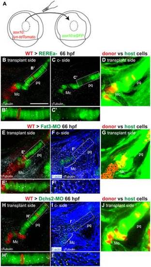
(A): presumptive CNC cells were transplanted from sox10:lyn-tdTomato- to sox10:eGFP transgenic embryos at the shield stage (B–D): WT transplants rescue cartilage stacking and polarity non-cell autonomously in rerea-/- embryos. Embryos stained for anti-acetylated tubulin (white). (B, B′) single confocal slice showing rescue of stacking and polarity in (rerea-/-; sox10:eGFP) mutant cells by a (WT; sox10:lyn-tdTomato) transplant. (C, C′) contralateral side without transplant. (D) Transplanted side with increased brightness in the green and red channels to show lineal contributions in non-cartilage cells. (E–J) Non cell-autonomous rescue of cartilage differentiation, stacking and polarity in (Fat3-MO; sox10:eGFP)(E, E′) or (Dchs2-MO; sox10:eGFP)(H, H′) embryos by (WT; sox10:lyn-tdTomato) transplants. (F, F′, I, I′) contralateral sides without transplants, stained for cortical actin with phalloidin (blue) to reveal cell outlines. (G, J) Transplanted side images with increased brightness in the green and red channels to show lineal contributions in non-cartilage cells. Scale bar = 50µm. Mc: Meckel′s; pq: palatoquadrate. |