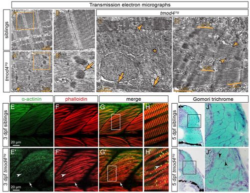
Cytoplasmic rod formation in tmod4trg. (A) Transmission electron micrographs of skeletal muscle from siblings at 3 dpf display the typical myofibril striation and well-aligned sarcomeres. (A′) In contrast, the myofibrils of tmod4trg mutants appear non-uniform, and filaments are misoriented. Sarcomere H-zones are less defined (arrowhead) and Z-disks are widened with electron-dense inclusions of various sizes and shapes. (B,B′) Magnified views of the respective boxes in A and A′ reveal the lattice pattern of the cytoplasmic rods (arrow) that is typical for nemaline rods of individuals with NM. (C) In addition to abnormal sarcomeres (arrowhead), organised sarcomeres (arrows) rarely form in tmod4trg, and filaments are often scattered throughout myofibres (asterisk). (D) Brackets mark the various lengths of thin filaments of tmod4trg from short (0.60 μm) to long (0.75 μm). In organised myofibrils, thin filaments are of lengths comparable to those of siblings (0.68 μm). Indistinct H-zones are marked by arrowheads. (E–H) At 3 dpf, labelling of F-actin with phalloidin (red) and actinin using an antibody (green) shows the typical myofibril striation in siblings. (G,H) Merged images, H shows magnification of the boxed area indicated in G. (E′-H′) In tmod4trg mutants, actinin and actin colocalize in cytoplasmic aggregations. In contrast to the internal fast myofibres that show abundant cytoplasmic rods (arrowheads), the superficial slow muscle fibres, which in zebrafish form a single layer on the lateral outline of the somites, do not display cytoplasmic rods (arrows). (G′,H′) Merged images, H′ shows magnification of the boxed area indicated in G′. (I,I′) On cross sections at 5 dpf, Gomori trichrome staining indicates the presence of cytoplasmic rods in tmod4trg that are reminiscent of nemaline rods. (J,J′) Magnifications of boxes indicated in I and I′, respectively. Arrowheads mark nemaline-like cytoplasmic rods.
|
