
Fig. 2
- ID
- ZDB-FIG-150203-18
- Publication
- Ito-Amano et al., 2014 - Temporal and spatial expression patterns of bone morphogenetic protein 3 in developing zebrafish
- Other Figures
- All Figure Page
- Back to All Figure Page
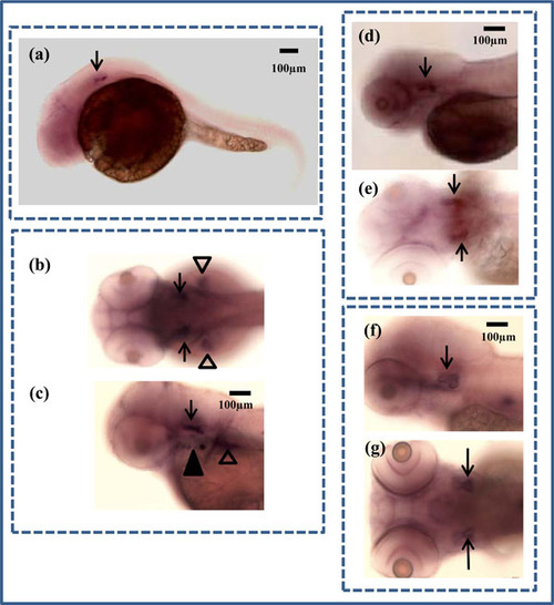
In situ hybridization of zbmp3 in developing zebrafish. (a) Lateral view of a 1 dpf zebrafish. Arrow indicates otic vesicle. (b) Dorsal view of a 2 dpf zebrafish. Arrows indicate otic vesicles and arrowheads show pectoral fins. (c) Lateral view of a 2 dpf zebrafish. Arrow indicates otic vesicle, black arrowhead shows opercle and branchiostegal ray, and white arrowhead shows pectoral fin. (d) Lateral view of a 3 dpf zebrafish. Arrow indicates otic vesicle. (e) Dorsal view of a 3 dpf zebrafish. Arrows indicate otic vesicles. (f) Lateral view of a 5 dpf zebrafish. Arrow indicates otic vesicle. (g) Dorsal view of a 5 dpf zebrafish. Arrows indicate otic vesicles. |
| Gene: | |
|---|---|
| Fish: | |
| Anatomical Terms: | |
| Stage Range: | Prim-5 to Day 5 |