
Fig. 1
- ID
- ZDB-FIG-150129-1
- Publication
- Zhang et al., 2014 - Identification of Annexin A4 as a hepatopancreas factor involved in liver cell survival
- Other Figures
- All Figure Page
- Back to All Figure Page
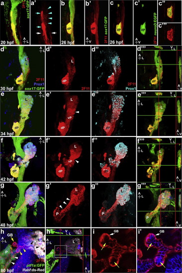
2F11 mAb labels hepatopancreas progenitors during organogenesis. (a) and (a′) Ventral view of a 20 hpf Tg(sox17:GFP) foregut showing broad low 2F11 labeling (turquoise arrowheads), and higher labeling in dorsal pancreas cells (turquoise arrows) and the pronephric ducts (white arrows). (b) and (c′) Lateral view of 26 hpf Tg(sox17:GFP) (b) and Tg(neuroD:EGFP) (c) foregut showing 2F11 labels the dorsal pancreas (dP) with a single focal plane shown in (c′′) and (c′′′). (d)-(g′′′) Ventral view of the Tg(sox17:GFP) foregut with Prox1 and 2F11 labeling at 30, 34, 42 and 48 hpf; (d′′′), (e′′′), (f′′′), (g′′′) digital sections along X, Y, and Z axes showing higher 2F11 staining on the ventral side of the endoderm (green lines indicate the positions of the X-axis sections, right insets; red lines indicate the positions of the Y-axis sections, top insets, a larger image of (f′′′) is shown in Fig. S1). (d)-(d′′) 2F11 staining increases in the Prox1+ budding liver (L) at 30 hpf. (e)-(e′′) At 34 hpf, 2F11 labeling increases in the Prox1+ ventral pancreas (vP) with broad cellular localization. Note the elevated staining in cells between the liver and ventral pancreas (white arrowhead). (f) and (f′′) At 42 hpf, 2F11 labeling increases further in the cells between liver and ventral pancreas with broad cellular localization. (g) and (g′′) At 48 hpf, 2F11 labels the entire hepatopancreas system with elevated staining and non-nuclear localization (white arrowheads) in cells between the liver and pancreas (P). (h) Ventral view of the Tg(ptf1a:GFP);Tg(lfabf:ds-Red) foregut at 80 hpf showing 2F11 labeling becomes restricted to the gallbladder (GB; yellow arrow) and ducts, including intrapancreatic ducts (white arrowheads), intrahepatic ducts (black arrowheads), and extra hepatopancreatic ducts (yellow arrowheads). (h′) Digital sections along X, Y, and Z axes showing higher 2F11 staining in the ventral region of the ducts. (i) and (i′) Higher magnification of (h′) (white square inset) showing 2F11 basal-lateral cellular localization in ductal columnar epithelium where it does not overlap with apically restricted Cadherin (yellow arrows). |
| Genes: | |
|---|---|
| Antibody: | |
| Fish: | |
| Anatomical Terms: | |
| Stage Range: | 20-25 somites to Protruding-mouth |
Reprinted from Developmental Biology, 395(1), Zhang, D., Golubkov, V.S., Han, W., Correa, R.G., Zhou, Y., Lee, S., Strongin, A.Y., Dong, P.D., Identification of Annexin A4 as a hepatopancreas factor involved in liver cell survival, 96-110, Copyright (2014) with permission from Elsevier. Full text @ Dev. Biol.