
Fig. 6
- ID
- ZDB-FIG-150115-7
- Publication
- Hartsock et al., 2014 - In vivo analysis of Hyaloid vasculature morphogenesis in zebrafish: A role for the lens in maturation and maintenance of the Hyaloid
- Other Figures
- All Figure Page
- Back to All Figure Page
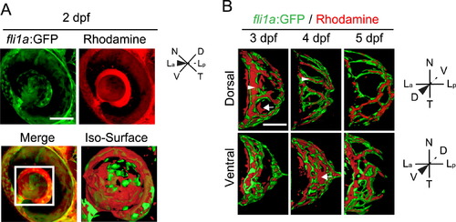
Microangiography demonstrates that the hyaloid vessel is fully enclosed by 5 dpf. (A) Maximum projection images of the eye of a fli1a:GFP (green) embryo injected with 50 kDa rhodamine dextran (red). Merged image highlights that rhodamine is detected throughout the vitreous. Iso-surface rendering of merged image. (B-D) Iso-surface renderings of maximum projections collected from the eyes of 3-5 dpf fli1a:GFP (green) embryos injected with 50 kDa rhodamine dextran (red) and imaged either dorsally or ventrally. Arrowhead highlights rhodamine containment in a fli1a:GFP vessel and arrows highlight rhodamine outside of vessels. hh:mm. Scale bars=50 µm. D: Dorsal, V: Ventral, N: Nasal, T: Temporal, La: Lens anterior, Lp: Lens posterior. |
| Gene: | |
|---|---|
| Fish: | |
| Anatomical Term: | |
| Stage Range: | Long-pec to Day 5 |
Reprinted from Developmental Biology, 394(2), Hartsock, A., Lee, C., Arnold, V., Gross, J.M., In vivo analysis of Hyaloid vasculature morphogenesis in zebrafish: A role for the lens in maturation and maintenance of the Hyaloid, 327-39, Copyright (2014) with permission from Elsevier. Full text @ Dev. Biol.