
Fig. 6
- ID
- ZDB-FIG-141223-10
- Publication
- Shimizu et al., 2014 - Hipk2 and PP1c Cooperate to Maintain Dvl Protein Levels Required for Wnt Signal Transduction
- Other Figures
- All Figure Page
- Back to All Figure Page
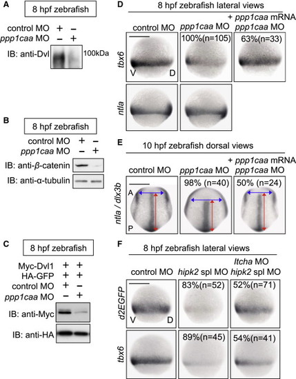
In Zebrafish, PP1c Cooperates with Hipk2 to Sustain Wnt Signal Transduction while Itch Counteracts Hipk2 Activity (A–C) PP1c is required for the protein stability of endogenous Dvl (A) and β-catenin (B) and exogenous mouse Dvl1 (C) in zebrafish. Zebrafish embryos were injected with MOs without (A and B) or with (C) mouse Myc-Dvl1 and HA-GFP mRNA. Extracts were harvested from the embryos at 8 hpf. In (A), embryo extracts were immunoprecipitated and then immunoblotted with anti-Dvl. In (B) and (C), extracts were immunoblotted with indicated antibodies. (D and E) PP1c is required for tbx6 expression and CE. Embryos were injected with control MO or ppp1caa MO (2 ng in D, 4 ng in E) with or without MO-insensitive ppp1caa mRNA. Panels show whole-mount in situ hybridization of tbx6, ntla, or dlx3b in zebrafish embryos. (F) Itch counteracts Hipk2 activity in zebrafish. Embryos were injected with MOs as indicated. Panels show whole-mount in situ hybridization of OTM:d2EGFP (d2EGFP) or tbx6 in OTM:d2EGFP-transgenic (top) or nontransgenic (bottom) zebrafish embryos. In (D)–(F), the percentages of embryos showing similar expression patterns and the total number of MO-injected embryos (n) are shown under each image. |
| Genes: | |
|---|---|
| Fish: | |
| Knockdown Reagents: | |
| Anatomical Terms: | |
| Stage Range: | 75%-epiboly to Bud |
| Fish: | |
|---|---|
| Knockdown Reagents: | |
| Observed In: | |
| Stage: | Bud |