
Fig. 6
- ID
- ZDB-FIG-141007-67
- Publication
- Tryon et al., 2014 - Clonal Analysis of kit ligand a Functional Expression Reveals Lineage-Specific Competence to Promote Melanocyte Rescue in the Mutant Regenerating Caudal Fin
- Other Figures
- All Figure Page
- Back to All Figure Page
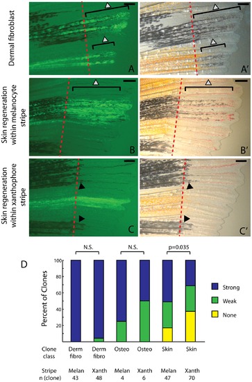
Skin clones show diminished ability to rescue melanocytes in xanthophores stripe regions of the regenerate. A-A′. Dermal fibroblasts strongly rescue melanocytes irrespective of where the clone occurs in the fin. B-C. Skin clones within the melanocyte stripe (B-B′) show more robust melanocyte rescue than skin clones that occur in a xanthophores stripe (C-C′). D. Dermal fibroblast, osteoblast, and skin clones were scored for the quality of melanocyte regeneration relative to their occurrence in a presumptive melanocyte stripe or xanthophore stripe. A single clone (as summarized in figure 4) may be scored as two clones if it occurs in both a xanthophore and melanocyte stripe region. Only skin clones showed a statistically significant difference in strength of rescue relative to the region in which the clone was regenerated (chi-squared 3×2 test, p value = 0.035.) Red dashed lines indicate the amputation plane. Grey arrowheads indicate newly differentiated kit-dependent melanocytes. Black arrowheads indicate previously differentiated kit-independent melanocytes. Scale bar = 250 mm. |