
FIGURE
Fig. 5
- ID
- ZDB-FIG-141007-66
- Publication
- Tryon et al., 2014 - Clonal Analysis of kit ligand a Functional Expression Reveals Lineage-Specific Competence to Promote Melanocyte Rescue in the Mutant Regenerating Caudal Fin
- Other Figures
- All Figure Page
- Back to All Figure Page
Fig. 5
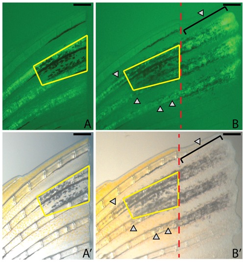
Expression of kitlga promotes differentiation of new melanocytes in the stump. A-A′. At the time of amputation and prior to induction of kitlga, melanocytes are well organized and form the proximal portion of the dorsal melanocyte stripe of the caudal fin (yellow trapezoid). B-B′. Following 7 days of kitlga expression, new melanocytes are visible both in the regenerate (brackets) as well as in the stump (grey arrowheads) in association with the dermal fibroblast clone. Red dashed lines indicate the amputation plane. Scale bar = 250 mm. |
Expression Data
Expression Detail
Antibody Labeling
Phenotype Data
Phenotype Detail
Acknowledgments
This image is the copyrighted work of the attributed author or publisher, and
ZFIN has permission only to display this image to its users.
Additional permissions should be obtained from the applicable author or publisher of the image.
Full text @ PLoS One