
FIGURE
Fig. 3
- ID
- ZDB-FIG-140909-22
- Publication
- Wang et al., 2014 - Grouper tshbeta Promoter-Driven Transgenic Zebrafish Marks Proximal Kidney Tubule Development
- Other Figures
- All Figure Page
- Back to All Figure Page
Fig. 3
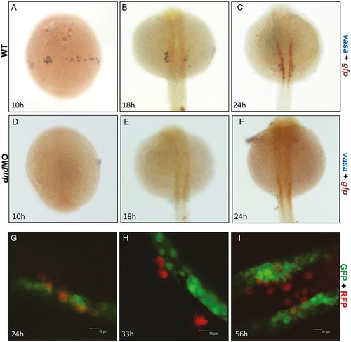
The gtshβ promoter-driven GFP was not localized with PGC. (A–C) dWISH of gfp (red) and vasa (purple) in Tg(gtshβ::GFP) embryos injecting control (A–C) and dnd-morpholino (dnd-MO) (D–F). (G–I) Tg(gtshβ::GFP) embryos injected with RFP-nos 32UTR mRNA (red) were observed using confocal microscopy at the indicated stages. Bar = 10 μm. |
Expression Data
Expression Detail
Antibody Labeling
Phenotype Data
Phenotype Detail
Acknowledgments
This image is the copyrighted work of the attributed author or publisher, and
ZFIN has permission only to display this image to its users.
Additional permissions should be obtained from the applicable author or publisher of the image.
Full text @ PLoS One