
Fig. 1
- ID
- ZDB-FIG-140908-11
- Publication
- Majumdar et al., 2000 - The zebrafish floating head mutant demonstrates podocytes play an important role in directing glomerular differentiation
- Other Figures
- All Figure Page
- Back to All Figure Page
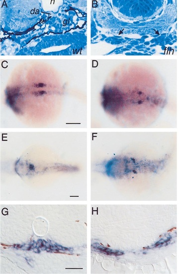
Glomerular development is altered in flh. Transverse histological sections (A, B). In wildtype zebrafish embryos at 2 days postfertilization (dpf) (A) a midline glomerulus (gl) is seen underneath the notochord (n) and dorsal aorta (da). A midline glomerulus is not seen in flh (B) at 2 dpf. Instead, clusters of cells are found in ectopic locations in flh (arrows in B). Whole-mount in situ hybridization with the wt1 probe (C-F). wt1 is expressed in nephric primordia in wildtype embryos at 20 h postfertilization (hpf) (C) and later at 48 hpf (E) in differentiated podocytes after pronephric primordia fuse in the midline. wt1 is expressed in flh nephric primordia at 20 hpf (D), but wt1-expressing cells remain in ectopic lateral positions at 48 hpf (F). Transverse sections of wt1 hybridized 36-hpf embryos (G, H). wt1-expressing cells are found at the midline in wildtype siblings (G) and in laterally positioned cells in flh (H). Some brown pigment granules are seen in these sections. Scale bars: (C, D) 200 µm; (E, F) 250 µm; (G, H) 50 µm. |
| Gene: | |
|---|---|
| Fish: | |
| Anatomical Terms: | |
| Stage Range: | 20-25 somites to Long-pec |
| Fish: | |
|---|---|
| Observed In: | |
| Stage Range: | Prim-25 to Long-pec |
Reprinted from Developmental Biology, 222(1), Majumdar, A. and Drummond, I.A., The zebrafish floating head mutant demonstrates podocytes play an important role in directing glomerular differentiation, 147-157, Copyright (2000) with permission from Elsevier. Full text @ Dev. Biol.