
Fig. 2
- ID
- ZDB-FIG-140812-19
- Publication
- Wiweger et al., 2014 - Possible effects of EXT2 on mesenchymal differentiation--lessons from the zebrafish
- Other Figures
- All Figure Page
- Back to All Figure Page
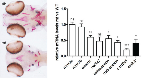
Bone development is impaired in the ext2-/-fish. A, Alizarin red stain for ossification in the craniofacial skeleton at 6dpf. Dermal bones: dentary (d), maxilla (mx), parasphenoid (p), endopterygoid (e), branchiostegal rays (br), opercle (op), cleithrum (cl), cartilage bones: hyomandibula (hm), ceratohyal (ch). ceratobranchial 5/pharyngeal arch (pa); and notochord (n), scale = 0.1 mm; B, The relative change in the expression of bone markers at 5dpf was evaluated by real time PCR and analysed by delta-delta-Ct in the homozygote ext2 mutants vs. wild type. The results represent an average from minimum four single embryos. Expression was normalized against slc25a5. Error bars indicate means with SEM. Expression of the ext2 was given as an example of a gene that was approximately 2-folds down-regulated and this under-expression was of biological relevance. |
| Fish: | |
|---|---|
| Observed In: | |
| Stage: | Day 6 |