
Fig. 1
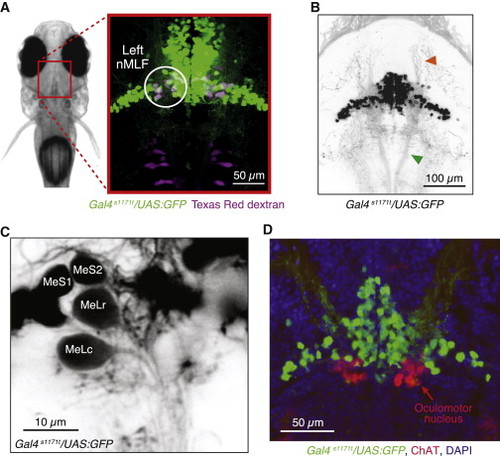
Enhancer Trap Line Gal4s1171t Drives Expression in the nMLF (A) Confocal projection of the midbrain in a 6-day-old Gal4s1171t/UAS:GFP fish. Optical sections (100 μm) were collapsed to yield a maximum intensity projection. Reticulospinal neurons were backfilled from the spinal cord with Texas Red dextran. Right panel is an expanded view of the nMLF region indicated by the red box in the left panel. Green cells are GFP-labeled by Gal4s1171t. Magenta cells are RS neurons in the rostral hindbrain and midbrain, labeled by Texas Red. White or pale magenta cells within the white circle are left nMLF neurons, which are double-labeled. (B) Confocal projection (80 μm) of the dorsal expression pattern in Gal4s1171t/UAS:GFP. The axon tract of the MLF (green arrowhead) and dendrites exiting the nMLF (orange arrowhead) are highlighted. (C) Two-photon image of a single plane highlighting four identified nMLF neurons, MeLr, and MeLc, plus the newly identified MeS1 and MeS2. (D) Confocal image projection of a cryostat section (25 μm) from Gal4s1171t/UAS:GFP stained with antibodies to GFP (green) and ChAT (red). Cell nuclei are labeled with a DNA dye (blue). See also Figure S1 and Movie S1. |