
Fig. 1
- ID
- ZDB-FIG-140801-1
- Publication
- Coxam et al., 2014 - Pkd1 Regulates Lymphatic Vascular Morphogenesis during Development
- Other Figures
- All Figure Page
- Back to All Figure Page
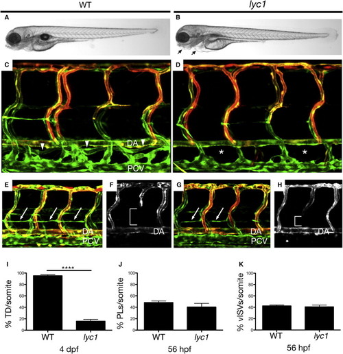
lyc1 Mutants Display Reduced Lymphatic Development (A and B) Overall morphology of wild-type siblings (A) and lyc1 mutants (B) at 4 dpf. (C and D) The vasculature Tg(fli1a:EGFPy1; flt1:tomatohu5333Tg) of (C) wild-type (WT) (arrowheads indicate thoracic duct) and (D) lyc1 mutants at 4 dpf (asterisks indicate absence of thoracic duct). (E and G) The vasculature Tg(fli1a:EGFPy1; flt1:tomatohu5333Tg) in wild-type sibling (E) and mutant embryos (G) at 56 hpf (arrows indicate lymphatic precursors known as parachordal lymphangioblasts, PLs). (F and H) flt1:tomatohu5333Tg expression marks the arterial ECs, a loss of signal (brackets) indicating venous intersegmental vessels (vISVs). (I–K) Quantification of (I) thoracic duct extent across ten somites (WT n = 40, lyc1 n = 17), (J) parachordal lymphangioblasts (WT n = 78, lyc1 n = 17), and (K) venous sprouts (WT n = 40, lyc1 n = 15). DA, dorsal aorta; PCV, posterior cardinal vein. Error bars indicate SEM. See also Figures S1 and S2. |
| Genes: | |
|---|---|
| Fish: | |
| Anatomical Terms: | |
| Stage Range: | Long-pec to Day 4 |
| Fish: | |
|---|---|
| Observed In: | |
| Stage: | Day 4 |