
Fig. S4
- ID
- ZDB-FIG-140626-16
- Publication
- Okigawa et al., 2014 - Different combinations of Notch ligands and receptors regulate V2 interneuron progenitor proliferation and V2a/V2b cell fate determination
- Other Figures
- All Figure Page
- Back to All Figure Page
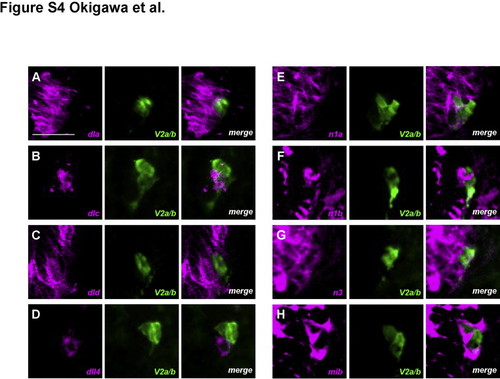
Expression patterns of Notch ligands and receptors in and around V2 neuronal lineage cells. deltaA, deltaD, notch1a, notch1b, notch3, and mib mRNAs were detected in the spinal cord and in and around V2 neurons, visualized with an anti-GFP antibody (A, C, E, F, G, and H). In contrast, deltaC was expressed in one of a pair of V2 neurons, which was likely to be a V2a cell (B). dll4 was detected not in but adjacent to V2 neurons (D). Transverse sections through the trunk region in embryos at 24 hpf. All images are of the right lower portion of the neural tube. mRNAs were mainly in the cytoplasm, whereas GFP was in both the nucleus and cytoplasm. Bar scale: 20 µm. |
Reprinted from Developmental Biology, 391(2), Okigawa, S., Mizoguchi, T., Okano, M., Tanaka, H., Isoda, M., Jiang, Y.J., Suster, M., Higashijima, S.I., Kawakami, K., Itoh, M., Different combinations of Notch ligands and receptors regulate V2 interneuron progenitor proliferation and V2a/V2b cell fate determination, 196-206, Copyright (2014) with permission from Elsevier. Full text @ Dev. Biol.