
Fig. 1
- ID
- ZDB-FIG-140624-27
- Publication
- Zeng et al., 2014 - Cadm4 Restricts the Production of Cardiac Outflow Tract Progenitor Cells
- Other Figures
- All Figure Page
- Back to All Figure Page
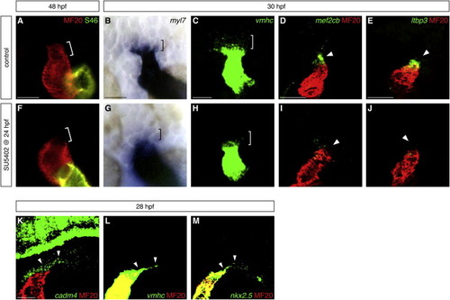
Fgf Signaling Promotes Formation of OFT Progenitor Cells (A and F) Hearts at 48 hpf, frontal views, arterial pole up, stained with MF20 (red) and S46 (green) antibodies to visualize the OFT (red, bracket), ventricle (red), and atrium (yellow). The OFT is morphologically evident in control DMSO-treated embryos (A) and absent in embryos treated with 10 μM SU5402 from 24 to 48 hpf (F). (B–E and G–J) In situ hybridization highlights the presumed OFT progenitor cells adjacent to the arterial pole at 30 hpf. Dorsal views (B, C, G, and H) and lateral views (D, E, I, and J), arterial pole up; (C–E and H–J) partial reconstructions of confocal z stacks. The presumed OFT progenitors (brackets) express low levels of myl7 (blue, B) and vmhc (green, C); very few of these cells are found in embryos treated with SU5402 from 24 to 30 hpf (G and H). Expression of mef2cb (green, D) and ltbp3 (green, E) is visible in the progenitor cells (arrowheads) residing next to the differentiated myocardium of the heart tube (MF20, red); this population is barely detectable in SU5402-treated embryos (I and J). (K–M) Fluorescent in situ hybridization shows that cadm4 (green, K) is expressed in the region where OFT progenitor cells are thought to reside (arrowheads), as well as in neuronal tissues as previously reported ( Pietri et al., 2008). vmhc (green, L) and nkx2.5 (green, M) are expressed at low levels within the same region (arrowheads), as well as at high levels throughout the heart tube. Differentiated myocardium is marked by MF20 (red); partial reconstructions of lateral views at 28 hpf. Scale bars, 50 μm. See also Figure S1 and Table S1. |
| Genes: | |
|---|---|
| Antibodies: | |
| Fish: | |
| Condition: | |
| Anatomical Terms: | |
| Stage Range: | Prim-5 to Long-pec |
| Fish: | |
|---|---|
| Condition: | |
| Observed In: | |
| Stage: | Long-pec |