
Fig. 3
- ID
- ZDB-FIG-140624-14
- Publication
- Kelly et al., 2014 - A Pak1/Erk Signaling Module Acts through Gata6 to Regulate Cardiovascular Development in Zebrafish
- Other Figures
- All Figure Page
- Back to All Figure Page
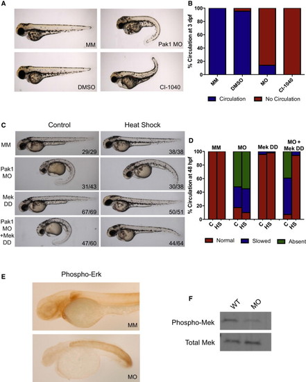
Pak1 Signals through the Erk Pathway in Heart Development (A) Comparison of chemical inhibition of Mek and pak1 morphants at 3 dpf. WT embryos were placed in egg water containing dimethyl sulfoxide (DMSO) or 0.5 µM CI-1040 at the one-cell stage. The water was changed every 24 hr with new drug. The embryos were analyzed for gross morphology and presence or absence of circulation. (B) Quantification of circulation seen with Mek inhibition at 48 hpf. (C) Representative images of 48 hpf pak1 morphants rescued by an active form of Mek (Mek DD). An inducible Mek1 DD expression plasmid was coinjected with the pak1 MO at the one-cell stage, followed by heat shock at 24 hpf as indicated. (D) Quantification of circulation seen with the addition of Mek DD. (E) Immunohistochemistry for phosphorylated Erk. (F) Immunoblot for phosphorylated Mek in whole embryos injected with either MM or pak1 MO. |
| Antibody: | |
|---|---|
| Fish: | |
| Knockdown Reagent: | |
| Anatomical Terms: | |
| Stage: | Prim-5 |
| Fish: | |
|---|---|
| Condition: | |
| Knockdown Reagent: | |
| Observed In: | |
| Stage Range: | Long-pec to Protruding-mouth |
Reprinted from Developmental Cell, 29, Kelly, M.L., Astsaturov, A., Rhodes, J., Chernoff, J., A Pak1/Erk Signaling Module Acts through Gata6 to Regulate Cardiovascular Development in Zebrafish, 350-9, Copyright (2014) with permission from Elsevier. Full text @ Dev. Cell