
Fig. 4
- ID
- ZDB-FIG-140623-18
- Publication
- Thomas-Jinu et al., 2013 - Dynamic expression of neurexophilin1 during zebrafish embryonic development
- Other Figures
- All Figure Page
- Back to All Figure Page
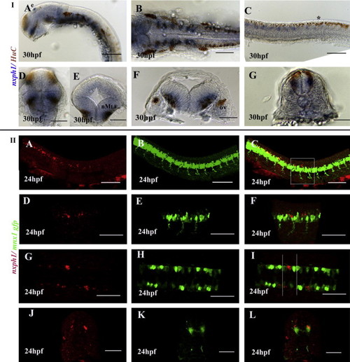
nxph1 is expressed in neurons, mostly interneurons. (I) Anti-HuC staining for post-mitotic neurons in 30 hpf embryos stained for nxph1 (A–G). Lateral view of zebrafish brain (A), frontal view of forebrain (D), transverse view of midbrain (E), dorsal (B) and transverse (F) view of hindbrain, lateral (C) and transverse (G) view of spinal cord at 36 hpf. Abbreviations: nMLF, nuclei of the media longitudinal fascicle); ov, otic vescicle; e, epiphysis. Scale bar: A–C (40 µm) and D–G (20 µm). (II) Lateral (A–F), dorsal view (G–I) and transverse section of spinal cord (J–L) of Tg(mnx1:GFP) zebrafish at 24 hpf, anterior to the left. Scale bar: A–C, J–L (40 µm) and D–I (20 ¼m). nxph1 is expressed ventral (blue) to the Rohon-Beard sensory neurons () stained by HuC (I) and dorsal (red) to the motor neurons marked by Tg(mnx1:GFP)(II), thus indicating its expression in the interneurons. |
| Genes: | |
|---|---|
| Fish: | |
| Anatomical Terms: | |
| Stage Range: | Prim-5 to Prim-15 |
Reprinted from Gene expression patterns : GEP, 13(8), Thomas-Jinu, S., and Houart, C., Dynamic expression of neurexophilin1 during zebrafish embryonic development, 395-401, Copyright (2013) with permission from Elsevier. Full text @ Gene Expr. Patterns