
Fig. S2
- ID
- ZDB-FIG-140619-8
- Publication
- Goetz et al., 2014 - Endothelial cilia mediate low flow sensing during zebrafish vascular development
- Other Figures
- All Figure Page
- Back to All Figure Page
|
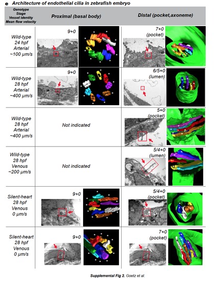
a. CLEM and electron tomography analysis of the venous cilium presented in Fig.2b,c,e. The cilium is located in the caudal vein of a 28 hpf embryo. A single tomogram spanning the celllumen interface is presented. Daughter and mother centrioles are in red, interconnecting fibers are in white, transition fibers are in yellow, plasma membrane is in green, microtubules are in red. The cilium contains five microtubules in the axoneme. b-d. CLEM analysis of the arterial cilium presented in Fig.2. b. Single TEM image of the region containing the cilium of interest (green boxed region, endothelium is in red, blood cells in blue). c. 3D modeling of the region of interest based on all the TEM micrographs containing the cilium of interest (d.) Note that the deflection angle of the cilium is conserved upon sample processing. See also Movie 10. (CA=Caudal Artery). e. Table showing al the cilia analyzed by electron tomography classified by genotype, stage, and vessel identity. Microtubule content, TEM images and 3D models are presented for both the basal body (proximal) and axoneme (distal) region of the cilia. Note the decrease in microtubule content in all the studied cilia. 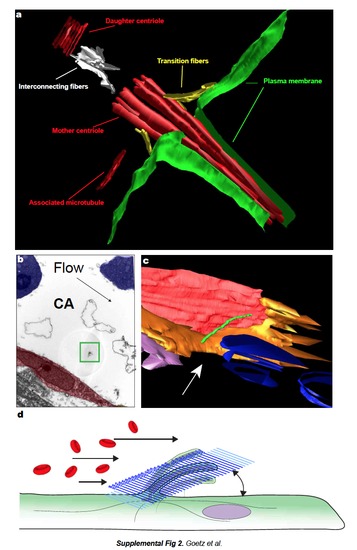
|