|
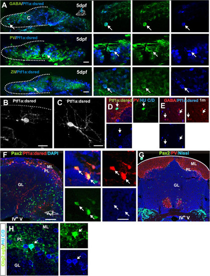
Cerebellar progenitors gives rise to distinct cell lineages. (A) Co-localization (arrow) of Ptf1a:DsRed (blue) and GABA, PV or ZII (green) in inhibitory neurons in the cerebellum of a 5-day-old larvae; (B,C) Ptf1a:DsRed+ cells showing morphologies of differentiating stellate and Golgi neurons; (D) A PV- (red) Ptf1a:DsRed+ (green) and HU C/D+ (blue) stellate cell in the ML; (E) Co-localization (arrows) of Ptf1a:DsRed (blue) and GABA (red) cells in the cerebellum of a 1-month-old juvenile zebrafish; (F) Cross section of the juvenile cerebellum showing abundant co-localization (white arrow) of Ptf1a:DsRed (red) and Pax2 (green); (G) Overview of a cross section of the adult cerebellum showing Pax2 (green) labeled Golgi neurons in the GL and PL. Purkinje neurons in the PL are labeled with parvalbumin (red); (H) Co-localization (arrow) of Olig2:egfp (green) and HU C/D (blue) cell in the PL of an adult zebrafish.
|