
Fig. 1
- ID
- ZDB-FIG-140527-8
- Publication
- Steiner et al., 2014 - Dynamic gene expression by putative hair-cell progenitors during regeneration in the zebrafish lateral line
- Other Figures
- All Figure Page
- Back to All Figure Page
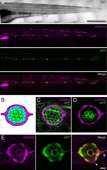
Expression of fluorescent proteins in mantle cells of the posterior lateral-line system. (A) Confocal mosaic images of a living alpl:mCherry;Et20 larva at 4 days postfertilization (dpf) demonstrate expression of mCherry (magenta) overlapping with that of GFP (green) in neuromasts and interneuromast cells. (Scale bar: 500 μm.) The same color code applies in C–E. (B) A neuromast comprises at least three cell types: hair cells (green), supporting cells (aqua), and mantle cells (magenta). (C) mCherry expression in alpl:mCherry larvae is limited to a subset of cells at the periphery of the neuromast. The image represents a confocal slice through a living alpl:mCherry; Tg(8.0cldnb:lynEGFP)zf106 larva, in which all cells of the neuromast express membrane-tethered GFP. (D) An alpl:mCherry;pou4f3:GFP animal expresses mCherry in peripheral cells, but that marker is excluded from hair cells that express membrane-tethered GFP instead. (E) mCherry and GFP have extensively overlapping but not identical expression patterns in mantle cells of alpl:mCherry;Et20 larvae. The arrowheads indicate two mCh+, GFP cells. (Scale bar: C–E, 10 μm.) |
| Genes: | |
|---|---|
| Fish: | |
| Anatomical Terms: | |
| Stage: | Day 4 |