FIGURE

Fig. 7

ID
ZDB-FIG-140321-26
Publication
Chen et al., 2001 - Analysis of the zebrafish smoothened mutant reveals conserved and divergent functions of hedgehog activity
Other Figures
All Figure Page
Back to All Figure Page
Fig. 7

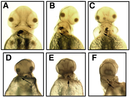
Hh activity and heart looping. All are ventral views of MF- 20-stained embryos at 48 hpf. Ventricle is stained darker than aorta. Anterior points upwards. Arrows indicate looping direction. (A-C) Despite morphological abnormalities, the heart of mutants (B,C) always loops to the right, as in wild-type embryos (A). (D-F) Inhibition of Hh activity by cyclopamine during gastrulation does not randomize heart looping. Morphology of the hearts are severely affected by the treatment in both wild-type (D,E) and mutants (F). Occasionally, the hearts of some treated embryos do not loop (E). However, reversal of looping has not been observed.

Expression Data
Antibody:
Fish:
Condition:
Anatomical Term:
Stage: Long-pec

Expression Detail
Antibody Labeling
Phenotype Data
Fish:
Condition:
Observed In:
Stage: Long-pec

Phenotype Detail
Acknowledgments
This image is the copyrighted work of the attributed author or publisher, and ZFIN has permission only to display this image to its users. Additional permissions should be obtained from the applicable author or publisher of the image. Full text @ Development