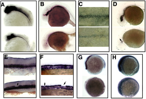
MFP development is independent of Hh activity in zebrafish. (A,B) Lateral view of wildtype and mutant embryos at 24 hpf showing FP markers such as shh (A) and axial (B) are present at reduced levels in smo embryos. (C) Close up dorsal view of axial expression in embryos at 24 hpf, indicating the lack of LFP and the presence of MFP in the mutant. (D) Expression of axial is markedly reduced in mutant embryos at 35 hpf. Axial can be seen in a small region in the ventral midbrain (arrow). (E) Lateral view of the expression of the MFP marker type II collagen 1a (col2a1) at 30 hpf indicating the fragmentation of MFP in the mutant (arrow). (F) Lateral view of col2a1 expression at 24 hpf indicating that 100 mM cyclopamine treatment from 4 to 10 hpf accelerated gap formation but did not eliminate the development of MFP. Arrows indicate gaps. (G) Side view of ptc1 expression in wild embryos at 10 hpf with (bottom) or without (top) 100 mM cyclopamine treatment showing that the treatment completely eliminates Hh activity. (H) Side view of ptc1 expression in the same batches of embryos as in (G) but sampled 2 hours after washing off cyclopamine, which indicates the inhibition is reversible.
|
