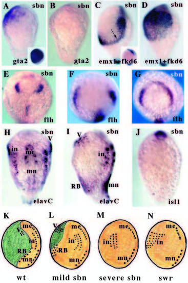
Sbn affects marginal cells fates and patterns of neurogenesis. All panels show 1-2 somite stage embryos. (A,B) Lateral views of gta2 expression (prospective epidermis) in mild (A) and severe (B) sbn- embryos. The inset panel shows expression in a wild-type embryo. Expression is retained in the anterior ventral region of the mildly affected embryo. (C,D) Lateral views of emx1 (prospective telencephalon) and fkd6 (prospective neural crest) expression in mild (C) and severe (D) sbn- embryos. The inset panel shows expression in a wild-type embryo. In the mildly affected embryo, expression is relatively normal in rostral regions, consistent with retention of gta2 expression (see A) in ventral ectoderm. However, more caudally, fkd6 expression is radialised (arrow). In the severely affected embryo, emx1 and fkd6 are expanded throughout the ventral ectoderm consistent with loss of gta2 in this region (see B). (E-G) Animal pole views of flh expression (prospective epiphysis) in progressively more severe sbn- embryos. flh expression is variably expanded in the sbn- embryos (see Fig. 2E,F for wild-type and swr- expression). (H) Dorsal and (I) lateral views of elavC expression in a sbn- embryo. Wild-type pattern of expression is shown in Fig. 3A,B. In these mildly affected sbn- embryos, interneurons are slightly expanded and dorsal sensory neurons are broadly expanded on the ventral side of the embryo. (J) Ventral view of isl1 expression in a severely affected sbn- embryo. Trigeminal and Rohon-Beard neurons are absent. (K-N) Summary schematics of patterns of neurogenesis in wild-type, sbn- and swr- embryos. In the mildly affected sbn- embryo, some non-neural ectoderm is retained and trigeminal and RB neurons are expanded on the ventral side of the embryo. In the severe sbn- embryo, trigeminal and RB neurons are lost and in the swr- embryo, interneurons are expanded throughout the ventral ectoderm. in, interneurons; me, medial neurons; mn, motor neurons; RB, Rohon-Beard neurons; V, trigeminal neurons.
|
