
Fig. 4
- ID
- ZDB-FIG-140307-33
- Publication
- Nasevicius et al., 1998 - Evidence for a fizzled-mediated wnt pathway required for zebrafish dorsal mesoderm formation
- Other Figures
- All Figure Page
- Back to All Figure Page
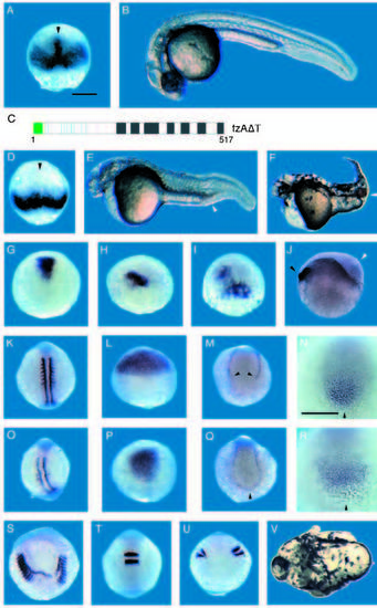
Misexpression of fzAΔT ventralizes zebrafish embryos. A,B,G,K,L,M,N and T are comparably staged control embryos. (C) Representation of fzAΔT, a dominant acting loss-of-function fz protein form that lacks the carboxy-terminal 60 amino acids (see Fig. 3). D-F, H-J, O-S and U,V show effects of fzAΔT misexpression. The dorsal mesoderm expression of chordin (arrowhead) is present in the control embryo (A) at approx. 65% epiboly but is missing in same stage fzAΔT-injected embryo (D). (E) 28-hour fzAΔT-injected embryo displaying a ventralized phenotype. Note extra cells in the tail (arrowhead) and reduced head structures. (F) Ventralized 2-day old fzAΔT-injected embryo. Note enlarged blood islands (arrowhead), a ventral-derived tissue. (G-J) Dorsal view of embryos stained for gsc. The stained region in fzAΔT-injected embryo (H) is reduced compared to control embryo (G). A significant fraction of fzAΔT-injected embryos showed mosaic gsc expression (I). (J) Lateral view of shield stage fzAΔT injected embryo, stained for gsc to mark the endogenous dorsoventral axis. Note thick cell layer (white arrowhead) observed during early gastrulation stages radially distant from the dorsal organizer (black arrowhead). (K,O) Dorsal view of mid-somitogenesis embryos stained for MyoD. Somitic mesoderm visualized by MyoD staining is reduced in fzAΔT-injected embryo (O) compared to control embryo (K); adaxial staining of MyoD is not significantly affected. (L,P) Dorsal view of 80% epiboly stage embryos stained for the anterior neuroectoderm marker otx-2. Note that otx-2 expression is significantly reduced in the fzAΔT-injected embryo (P) compared to control embryo (L). This phenotype is similar to the head reduction phenotype observed at 28 hours of development (E). (MN and Q-R). Ventral markers are expanded in fzAΔT-injected embryos, as judged by the tailbud expression of GATA-2 (M,Q) and eve-1 (N,R) in 10 somite embryos. In fzAΔT-injected embryos, ventral mesoderm stripes stained for GATA-2 are expanded and connected posteriorly (Q, black arrowhead) while in the uninjected embryo the stripes are separate (M, black arrowhead). eve-1 is expanded in fzAΔT-injected embryo (R) compared to control embryo (N). (S-V) High dose fzAΔT-injected embryos display a bifurcated axis phenotype distinct from effects on dorsoventral patterning (see text), which by 28 hours results in an embryo with only a single eye (V, white arrow indicates axis split). The phenotype can also be seen at the 10 somite stage when stained for MyoD (S) and Krox-20 (U). The stained areas are split into two distinct domains in fzAΔT-injected embryos compared to control embryos (K and T, accordingly). |