
Fig. 2
- ID
- ZDB-FIG-140306-5
- Publication
- Reifers et al., 1998 - Fgf8 is mutated in zebrafish acerebellar (ace) mutants and is required for maintenance of midbrain-hindbrain boundary development and somitogenesis
- Other Figures
- All Figure Page
- Back to All Figure Page
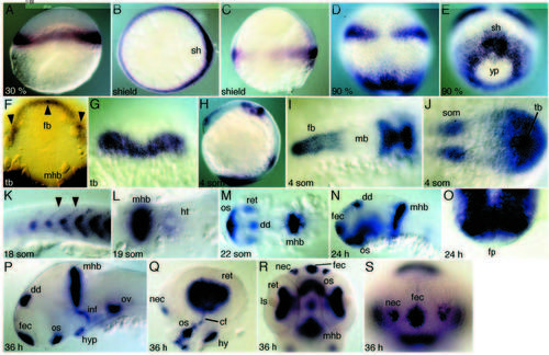
Expression of Fgf8 in wild-type embryos. (A) Fgf8 in the blastoderm margin at 30% epiboly. (B,C) At shield stage, Fgf8 is expressed in a dorsoventral gradient in the germ ring with a high point of expression in the shield (B, vegetal pole view; C, lateral view). (D,E) Graded expression persists in the margin of the blastoderm at 90% epiboly. Prospective anterior hindbrain expresses Fgf8 (D, dorsal view; slightly tilted in E). (F) Forebrain expression in a tailbud stage embryo (arrowheads point to high points of expression). (G) Prospective MHB domains fuse at the midline at tailbud stage. (H) 4-somite stage, lateral view. Expression in forebrain, mid-hindbrain region, segmental plate and tailbud. (I,J) Flat mount of H, depicting anterior and posterior expression domains. (K) Fgf8 expression at the anterior somite border (arrowheads). (L) Flat-mounted 19-somite embryo. Expression in the heart ring posterior to the MHB. (M) Flat mount at 22 somites; expression in the brain is detected at the MHB, dorsal diencephalon, retina and optic stalks. (N) Lateral view of a 24 hours embryo. Additional expression occurs in the facial ectoderm. (O) Thick cross section through the MHB demonstrating absence of expression in floorplate. (P) Lateral view of a dissected brain at 36 hours of development. Additional expression in the infundibulum, hypophysis and otic vesicle (eyes are removed). (Q) Details of expression in the retina, choroid fissure and the optic stalks. Additional expression is detected in nasal ectoderm and the hyoid. (R) Ventral view of head at 36 hours demonstrating expression in the retinal epithelium, but not in the lens. (S) Frontal view, expression in the facial and nasal ectoderm. cf, choroid fissure; dd, dorsal diencephalon; fb, forebrain; fec, facial ectoderm; fp, floorplate; ht, heart; hy, hyoid; hyp, hypophysis; inf, infundibulum; ls, lens; mb, midbrain; mhb, mid-hindbrain boundary; nec, nasal ectoderm; os, optic stalks; ov, otic vesicle; ret, retina; sh, shield; som, somites; tb, tailbud, yp, yolk plug. |