
Fig. 4
- ID
- ZDB-FIG-140305-21
- Publication
- Fürthauer et al., 1997 - A role for FGF-8 in the dorsoventral patterning of the zebrafish gastrula
- Other Figures
- All Figure Page
- Back to All Figure Page
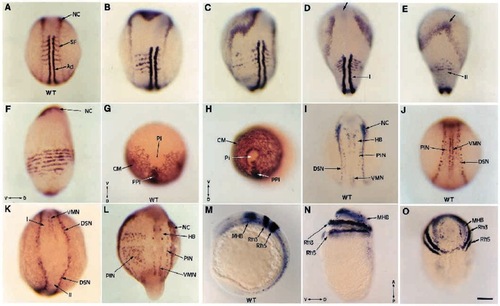
FGF-8 affects dorsoventral patterning of both mesoderm and ectoderm. After injection of FGF-8 RNA, embryos and their uninjected siblings were fixed at the beginning of somitogenesis, and probed using MyoD and AP2 in (A-F), snail2 (G,H), AS28 and AP2 (I-L), Krox20, Eng3 and AS28 in (M-O). Wild-type siblings in A,G,I,J,M). Labeling with MyoD and AP2 shows the gradual effects induced by FGF-8 from an enlarged paraxial territory (B) to a fragmented paraxial territory (C), or to the formation of a defective secondary axis on the ventral side of the embryo (E, compared to its dorsal wild-type side in D) or in extreme cases to a complete circularization of the paraxial territory (F). The cephalic mesoderm of these extreme dorsalized embryos, probed with snail2 (H compared to its wild-type control in G) is also circularized. The neural plate (the borders of which are labeled with AP2) is also expanded (B,C compared to A). When a secondary axis is formed (E), because of the absence of presumptive forebrain and midbrain, AP2 domain fuses anteriorly (arrow), while it remains separated in the primary axis (D). Analysis with AS28, Krox20 and Eng3 probes reveals the dorsolateralization effect of FGF-8 on the ectodermal layer. When a secondary axis (II) is formed as in K, dorsal sensory neurons (DSN) are present while ventral motor-neurons (VMN) are lacking. In embryos showing a dorsolateral expansion, as in L, the number of primary interneurons (PIN) on the affected side strongly increases (compared to the unaffected side or wild-type siblings in I,J). Strong dorsalized phenotypes (N,O) display a complete circularization of the neural plate visualized by Krox20 and Eng3 expression all around the yolk sac compared to wild-type control in (M). (A-D,I,J,L) Dorsal views; (E) ventral view; (G,H,O) animal pole views; (F,M,N) lateral views; (K) caudal view. (D,E) The dorsal and the ventral view of the same embryo. For references of the probes used, please see text. Ad, adaxial cells; CM, cephalic mesoderm; HB, hindbrain; MHB, midbrain-hindbrain boundary; NC, neural crest; Pi, pillow; PIN, primary interneurons; PPI, prechordal plate; Rh3, Rh5, rhombomeres 3 and 5; SF, somitic furrow; V, ventral; D, dorsal; A, anterior; P, posterior. Scale bar, 150 μm. |