|
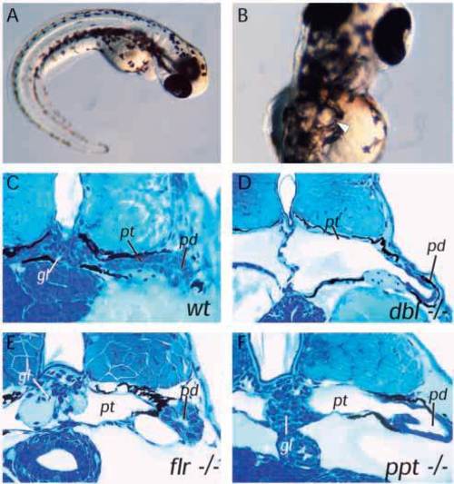
Cystic maldevelopment in zebrafish pronephric mutants. (A) The mutant double bubble (dbbm153) emerges from its chorion with a ventrally curved body axis and within 3-4 hours bilateral pronephric cysts (arrow in B) are evident. (C) Cross section of wildtype pronephros at 3.5 days pf. (D) Section of dbbm468 showing a grossly distended pronephric cyst in place of the pronephric tubule and a glomerulus reduced to a flattened septum at the midline. (E) The mutant fleer (flr) shows less severe cyst distension despite a flattened pronephric tubule epithelium and also a marked distension of the glomerular capillaries. (F) pao pao tang (pap) has an apparently intact glomerulus while the pronephric tubule epithelium is flattened and the pronephric duct is also distended. gl, glomerulus; pt, pronephric tubule; pd, pronephric duct.
|