
Fig. 3
- ID
- ZDB-FIG-140213-3
- Publication
- Wu et al., 2014 - Mediator subunit 12 coordinates intrinsic and extrinsic control of epithalamic development
- Other Figures
- All Figure Page
- Back to All Figure Page
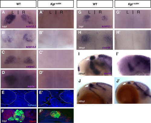
The med12/kgt mutant shows defects in habenular development. The habenular differentiation markers, kctd12.1, kctd12.2, nrp1a, and cadps2 were all absent in med12 mutants at 4 dpf (A-D′) and 2 dpf (G, G′), while the habenular cells were still present in the dorsal diencephalon in med12 mutants as shown by ToPro3 nuclear staining at 4 dpf (E, E′; habenular nuclei were outlined in white; the dark shadow on the left habenula in E′ is due to an overlying pigment cell). Fewer Elavl3-expressing neurons were observed (F, F′; at 2 dpf) and the expression of cxcr4b was significantly reduced in med12 mutants when compared to WT (H, H′; at 36 hpf). In med12 mutants, the presumptive habenular progenitor marker, dbx1b, was absent early (lateral views, at 28 hpf; I, I′) but reappeared in later stages (at 48 hpf, J, J′). |
| Genes: | |
|---|---|
| Antibody: | |
| Fish: | |
| Anatomical Terms: | |
| Stage Range: | Prim-5 to Day 4 |
| Fish: | |
|---|---|
| Observed In: | |
| Stage Range: | Prim-25 to Day 4 |
Reprinted from Developmental Biology, 385(1), Wu, S.Y., de Borsetti, N.H., Bain, E.J., Bulow, C.R., and Gamse, J.T., Mediator subunit 12 coordinates intrinsic and extrinsic control of epithalamic development, 13-22, Copyright (2014) with permission from Elsevier. Full text @ Dev. Biol.