
Fig. S6
- ID
- ZDB-FIG-140117-16
- Publication
- Nagashima et al., 2013 - A self-renewing division of zebrafish Muller glial cells generates neuronal progenitors that require N-cadherin to regenerate retinal neurons
- Other Figures
- All Figure Page
- Back to All Figure Page
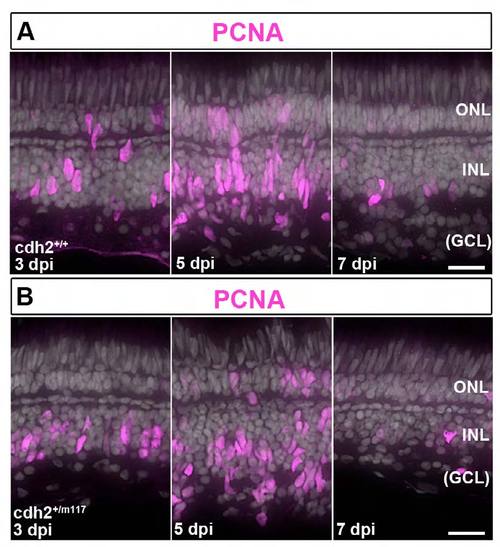
Injury-induced Müller glia produce proliferating retinal progenitors, but in cdh2+/m117 heterozygotes their morphology is abnormal after destruction of inner retinal neurons. (A,B) Immunocytochemistry for PCNA (magenta) in wild-type sib and cdh2+/m117 het retinas after intraocular injection of ouabain. (A) In the wild-type sib, PCNA is expressed in Müller glial nuclei that migrate apically at 3 dpi, and PCNA+ progenitors form elongated, spindle-shaped neurogenic clusters at 5 dpi. The number of PCNA+ cells decreases at 7 dpi. (B) In the cdh2+/m117 hets, at 5 dpi PCNA+ progenitors fail to form cohesive, neurogenic clusters, and their nuclei are rounded, rather than spindle-shaped. The number of PCNA+ cells is reduced at 7 dpi, as in wild-type sibs. Scales: 20 μm, A,B. |