FIGURE

Fig. S4

ID
ZDB-FIG-140117-14
Publication
Nagashima et al., 2013 - A self-renewing division of zebrafish Muller glial cells generates neuronal progenitors that require N-cadherin to regenerate retinal neurons
Other Figures
All Figure Page
Back to All Figure Page
Fig. S4

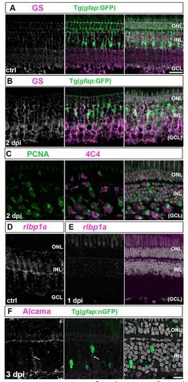
Basal processes of Muller glia collapse and markers of differentiation are downregulated after intraocular ouabain injection.
(A, B) Immunocytochemistry for the Müller glial marker glutamine synthetase (GS) (white/magenta) in unlesioned (A) and 2 dpi (B) gfap:GFP retinas with Müller glial reporter (green). GS+/GFP+ Müller glial processes in the inner retina are disrupted, whereas the organization of the outer retina, including the adherens junctions at the outer limiting membrane, remain intact at 2 dpi. (C) PCNA+ (green) activated microglia (4C4+, magenta) are abundant in the damaged inner retina at 2 dpi. (D, E) In situ hybridization for another Müller glia marker, rlbp1a (white/magenta, D), shows expression is gone at 1 dpi (E). (F) Weak Alcama immunoreactivity (magenta/ white) appears in the basal process (arrow) of a Müller glia with the inducible nGFP reporter (green) at 3 dpi. Scales: 20 μm, A-E; 10 μm, F.

Expression Data

Expression Detail
Antibody Labeling
Phenotype Data

Phenotype Detail
Acknowledgments
This image is the copyrighted work of the attributed author or publisher, and ZFIN has permission only to display this image to its users. Additional permissions should be obtained from the applicable author or publisher of the image. Full text @ Development