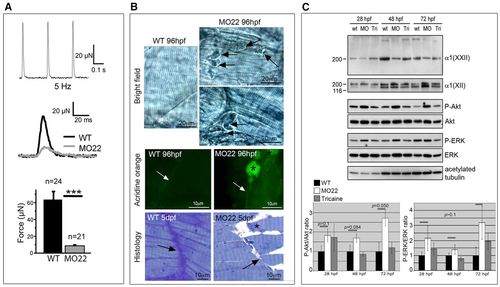
col22a1 knockdown leads to muscular dystrophic phenotype. (A) Recording of contraction in response to single supramaximal electric shocks applied to wild-type and MO-22-injected larvae. Twitch responses (middle panel) and mean contraction amplitudes (bottom panel) of 5 dpf wild-type (black) and MO22-injected (gray) larvae. Data are mean±s.e.m. ***P<0.001. (B) 96 hpf uninjected (WT) and MO22-injected (MO22) larvae. Light microscopy (top panel) revealed muscle detachment and retraction (MO22, arrows) from vertical myoseptum. Acridine Orange staining (middle panels): asterisks point to Acridine Orange-positive retracted muscle fibers in MO22 and arrows indicate myoseptum. Histology (bottom panels) of MO22 shows cell-free spaces, reduced MTJ interdigitations (arrows) and clear appearance of myosepta compared with WT. Asterisk indicates a detached and retracting muscle fiber. (C) Western blots with antibodies to phospho-Akt (P-Akt) and total Akt protein (Akt) or phospho-ERK1/2 (P-ERK1/2) and total ERK protein (ERK1/2) of extracts from uninjected (wt) and tricaine-treated (Tri) wild-type and MO22-injected (MO) embryos at different stages. Loading controls are antibodies to COLXII and acetylated tubulin. Data from one representative experiment are shown. Only one band is detected with ERK1/2 antibodies, as reported for zebrafish (Ng et al., 2012). Histograms show densitometric quantification. Phospho-protein levels were normalized against total protein levels. Data are mean±s.e.m. (n=3).
|
