
Fig. 1
- ID
- ZDB-FIG-140107-53
- Publication
- Borovina et al., 2013 - IFT88 Plays a Cilia- and PCP-Independent Role in Controlling Oriented Cell Divisions during Vertebrate Embryonic Development
- Other Figures
- All Figure Page
- Back to All Figure Page
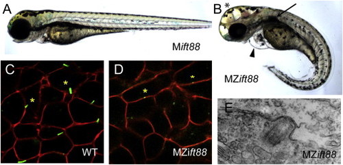
MZift88 Mutants Do Not Form Cilia (A and B) Lateral view of a 3-day-old (A) Mift88 embryo and (B) MZift88 mutant embryo exhibiting a curved body axis, pericardial edema (arrowhead), hydrocephalus (asterisk), and a cystic kidney (arrow). (C and D) Confocal images of (C) WT and (D) MZift88 mesoderm cells, at 9 hpf, expressing Arl13b-GFP (green) and memb-mRFP (red) revealed the presence of cilia on WT cells (asterisks, C). Only small puncta of Arl13b-GFP were visible on MZift88 mutant cells (asterisks, D). (E) Transmission electron microscopy micrograph of basal body docking in the neural tube of a 30hpf MZift88 mutant embryo. |
| Fish: | |
|---|---|
| Observed In: | |
| Stage Range: | 90%-epiboly to Protruding-mouth |