
Fig. S1
- ID
- ZDB-FIG-131211-48
- Publication
- Kwon et al., 2013 - The parallel growth of motoneuron axons with the dorsal aorta depends on Vegfc/Vegfr3 signaling in zebrafish
- Other Figures
- All Figure Page
- Back to All Figure Page
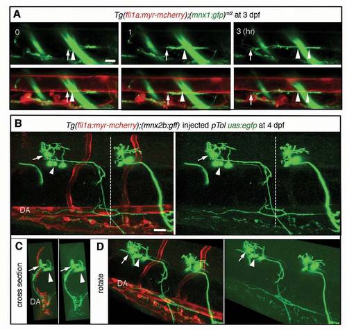
Motoneurons extend axons rostrally and caudally along the dorsal aorta. (A) Time-sequential confocal section images (lateral view) of an embryo expressing Tg(fli1a:myr mcherry);(mnx1:gfp)ml2. Elapsed time (hours) from the start point of time-lapse imaging (3 dpf) is shown in the upper panels. Upper panels, GFP images; lower panels, merged images of GFP and mCherry. Arrows indicate the tip of extending neuronal axon. Arrowheads denote the location of the tip when starting timelapse imaging. Note that axons grow rostrally along the dorsal aorta (DA). (B,C) 3D volume-rendered confocal stack images of the embryo indicated at the top in which a single cell was labeled. Left, the merged image of mCherry and GFP. Right, GFP image. Arrows and arrowheads indicate the different motoneuronal cell bodies. The 3D volume-rendered confocal stack images anterior to the dashed lines in B are shown in C. (D) Oblique views of B. Scale bars: 25 μm. |