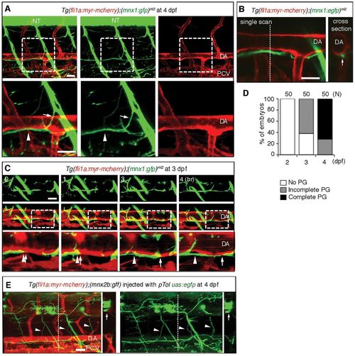
Axons of motoneurons develop beneath the dorsal aorta. (A) 3D-rendered confocal stack of fluorescence images (lateral view) of an Tg(fli1a;myr-mcherry);(mnx1:gfp)ml2 embryo at 4 dpf. Left column, merged images; center column, GFP images; right column, mCherry images. Lower panels are enlarged images of the boxed regions in the upper panels. Anterior is to the left. Arrows indicate the branch of the descending axon of motoneurons. Arrowheads indicate the extension of axons beneath the DA. (B) Left, a single scan confocal image from the stack shown in A. Right, a cross-section image of the stacked image shown in A at the level of the dashed line in the left panel. Arrow denotes the single fascicle of axons. (C) 3D-rendered confocal time-sequential stack images of an Tg (fli1a:myr-mcherry);(mnx1:gfp)ml2 embryo. Elapsed time (hours) from the start point of time-lapse imaging (3 dpf) is indicated. Top panels, GFP images; middle panels, merged images of GFP and mCherry; bottom panels, enlarged images of boxed regions of the middle panels. Arrows indicate the tip of a neuronal axon. Arrowheads denote the location of the tip when starting time-lapse imaging. (D) Parallel growth (PG) of motoneuron axons with the DA was quantitatively analyzed. ‘Complete’ indicates the complete continuity of the axon between the region above the rostral part of the yolk tube and that above the caudal part of the yolk tube. The number of embryos observed is indicated at the top. (E) Single-cell labeling experiment of an embryo of the indicated genotype. Left, 3D-rendered confocal merged image of mCherry and EGFP with a cross-section image at the level of the dashed line. Right, EGFP image with a cross-section image of the EGFP image at the level of the dashed line. Arrows and arrowheads denote the cell bodies of motoneurons in the neural tube and their axons extending beneath the DA, respectively. Scale bars: 25 μm. DA, dorsal aorta; NT, neural tube; PCV, posterior cardinal vein.
|
