
Fig. 1
- ID
- ZDB-FIG-131210-33
- Publication
- Hao et al., 2013 - Selective Small Molecule Targeting β-Catenin Function Discovered by In Vivo Chemical Genetic Screen
- Other Figures
- All Figure Page
- Back to All Figure Page
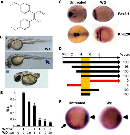
WD, which Dorsalizes Zebrafish Embryo, Inhibits Canonical Wnt Signaling (A) Chemical structure of WD, (Z) 3-chloro-2,3-bis(4-methoxyphenyl)acrylaldehyde, is shown. (B) WD dorsalizes zebrafish embryos. (i) Vehicle-treated WT zebrafish embryo at 48 hpf. (ii and iii) Dorsalized embryos treated with 10 μM WD. (i) Mildly dorsalized embryo is characterized by loss of ventral tail fin (arrow), whereas (ii) severely dorsalized embryo is characterized by severely shortened tail. (C) In situ hybridization of six-somite-stage (12 hpf) embryos was performed to evaluate expression of the dorsal markers Pax2.1, which marks the midhindbrain boundary, and Krox20, which marks rhombomeres 3 and 5. In comparison to untreated embryos (left), Pax2.1 and Krox20 expression are dramatically expanded in WD-treated embryos (right). Dorsal view, anterior is to the left. (D) Time window for dorsalization by WD (20 μM) is between 4 and 6 hpf. Numbers shown on right are percentage (%) of embryos dorsalized (dors) upon various WD treatment schemes. Left end of each bar or arrow represents the time when WD treatment was initiated, and the right end of each bar represents the time when WD was washed out (n > 100 embryos for each condition). (E) WD inhibited Wnt3-induced signaling in β-catenin-responsive TOPFLASH reporter assay (n = 4; results are represented as mean RLU [relative luciferase units] ± SE). (F) In situ hybridization for GFP expression in Wnt-dependent TOPFLASH-GFP transgenic zebrafish reveals that WD selectively abrogated Wnt signaling in the ventral and lateral regions (arrow) of the 50% epiboly stage (5.3 hpf) embryos, but not within the dorsal organizer (arrowhead). Ventral side is to the left. See also Figures S1, S2, and S3. |
| Genes: | |
|---|---|
| Fish: | |
| Condition: | |
| Anatomical Terms: | |
| Stage Range: | 50%-epiboly to 5-9 somites |
| Fish: | |
|---|---|
| Condition: | |
| Observed In: | |
| Stage Range: | 5-9 somites to Long-pec |