
FIGURE
Fig. S3
Fig. S3
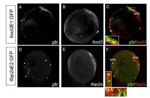
Double fluorescent ISH shows mosaic colocalization of enhancer:GFP with endogenous foxd3 and tfap2a mRNA expression. (A-F) Double fluorescent ISH for gfp (A) and foxd3 (B) in foxd3E1:GFP-injected embryos (merge in C, lateral view) and for gfp (D) and tfap2a (E) in tfap2aE2:GFP-injected embryos (merge in F, dorsal view) at 2-somites demonstrate mosaic colocalization of enhancer:GFP with the endogenous gene along the NPB (insets in C,F). (G) tfap2a and gfp also colocalize in the most anterior NPB as shown in a lateral magnification from a separate WT embryo. NPB, neural plate border; tb, tailbud. |
Expression Data
Expression Detail
Antibody Labeling
Phenotype Data
Phenotype Detail
Acknowledgments
This image is the copyrighted work of the attributed author or publisher, and
ZFIN has permission only to display this image to its users.
Additional permissions should be obtained from the applicable author or publisher of the image.
Full text @ Development