
Fig. 3
- ID
- ZDB-FIG-131015-5
- Publication
- Kwong et al., 2013 - The Role of Aquaporin and Tight Junction Proteins in the Regulation of Water Movement in Larval Zebrafish (Danio rerio)
- Other Figures
- All Figure Page
- Back to All Figure Page
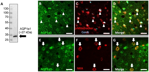
Aquaporin-1a1 is expressed in ionocytes on the skin of yolk sac in larval zebrafish. (A) A representative western blot showing that the eel aquaporin-1a1 (AQP1a1) antibody yielded a single immunoreactive band at ~27 kDa in lysates of 4 dpf zebrafish larvae. (B) Fluorescent immunohistochemistry and confocal microscopy revealed that the expression of AQP1a1 on the skin of yolk sac (green), and a subset of cells stained positively with Mitotracker® (red) and concanavalin-A (conA; blue). (D) is the merged images of B and C. Cells labelled with arrowheads represent areas of colocalization of AQP1a1 and Mitotracker® or conA. (E) Some AQP1a1-positive cells (green) were also colocalized with (F) Na+/K+-ATPase staining (NKA; red). (G) is the merged images of E and F. Cells labelled with arrows represent areas of colocalization of AQP1a1 and NKA. Scale bar = 25 μm. |
| Antibodies: | |
|---|---|
| Fish: | |
| Anatomical Term: | |
| Stage: | Day 4 |