
FIGURE
Fig. S8
Fig. S8
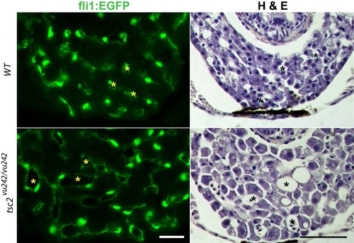
Increased size of blood vessels in the liver of tsc2 homozygous mutant zebrafish. The tsc2 mutant allele was crossed to transgenic fish expressing EGFP under the Fli1 promoter that directs expression in endothelial cells. Examples shown are liver from 7 day old larvae. Above images are wildtype (wt) control, below are tsc2 homozygous mutant sections. Left images are Fli1:EGFP, and right images hematoxylin and eosin staining. Asterisks (yellow in Fli1:EGFP, black in hematoxylin and eosin stain) denote the blood vessel lumen. Scale bar = 100 μm. |
Expression Data
Expression Detail
Antibody Labeling
Phenotype Data
| Fish: | |
|---|---|
| Observed In: | |
| Stage: | Days 7-13 |
Phenotype Detail
Acknowledgments
This image is the copyrighted work of the attributed author or publisher, and
ZFIN has permission only to display this image to its users.
Additional permissions should be obtained from the applicable author or publisher of the image.
Full text @ Dis. Model. Mech.