|
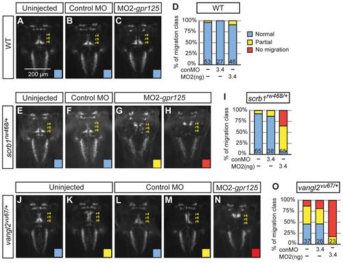
gpr125 interacts with scrb1/llk and vangl2/tri in FBMN migration. (A-C) Dorsal views of islet1(isl1):GFP-expressing neurons in uninjected, 3.4 ng control MO or 3.4 ng MO2-gpr125 injected wild-type siblings of scrb1/llk or vangl2/tri heterozygous embryos at 48 hpf. Anterior is upwards. r4 (rhombomere 4), r5 and r6 positions are labeled. (D) Frequency of FBMN migration phenotypic classes observed in wild-type embryos. Blue, normal; yellow, partial; red, no migration. (E-H) Dorsal views of isl1:GFP-expressing neurons in uninjected, 3.4 ng control MO or 3.4 ng MO2-gpr125-injected scrb1/llkrw468/+ embryos at 48 hpf. (I) Frequency of the FBMN migration phenotypic classes observed in scrb1/llkrw468/+ embryos. (J-N) Dorsal views of isl1:GFP-expressing neurons in uninjected, 3.4 ng control MO or 3.4 ng MO2-gpr125-injected vangl2/trivu67/+ embryos at 48 hpf. (O) Frequency of FBMN migration phenotypic classes observed in vangl2/trivu67/+ heterozygotes.
|